Case of 18 years old male patient with b/l limb weak ness and edema
G sai tejaswini
Roll no -61
Ihave been given this case to solve in an attempt to understand the topic of patient clinical data analysis to develop my competency in reading and comprehending clinical data including history, clinical findings, inveatigations and come up with diagnosis, treatment plan.
Entire real patient clinical problem in this link herehttps://srianugna.blogspot.com/2020/05/hello-everyone.html
A 18 years old male patient with bilateral lower limb weakness since 20 days the weak ness was started 2 years back in proximal region and gradualy progressive to distal region. bilateral non pitting type of edema in lower limbs. H/o difficulty of squating position and getting from tha postion. On examination there is areflexia.
Weakness cause may be nuero genic , myogenic, traumatic cause .
No h/o trauma so it is ruled out.
Neurogenic may be UMN LESION, LMN LESION. But the features are some related to lmn.
LOWER MOTOR LESION:
Anterior horn cells
Nerve roots
Peripheral nerves
Neuromuscular junction
Muscle
Anterior horn cells: it includes spinal muscular atrophy( areflexia, weakness,muscle twitching respiratory muscle involvement should be seen .diagnosis by gentic testing, ct , mri .By features , investigations it is ruled out.
Nerve root( radiculopathies): there should be pain, radiation of pain diagnosis by ct , mri. By above things we can rule out .
Peripheral nerve involvement ruled out by nerve conduction studies,.
Neuromuscular junction -by features and electromyography it can be ruled out from other diseses.
Muscle(myogenic causes): It may include
Inflammatory causes- muscle biopsy is done and impression given-no suggestive of polymyositis.
Metabolic causes -no features of glycogen storage disorders.
Drug induced-no history of drug intake.
Endocrine causes-thyroid profile is normal thyroid myopathy is ruled out.
Diagnosis may be muscular dystrophy (muscle biopsy)
Muscular dystrophy:
-myotonic dystrophy
- duchene's muscular dystrophy
--Beckers muscular dystrophy
Myotonic dystrophy :it involves facial muscle by sparing proximal muscles so it is ruled out.
Duchenes muscular dystrophy, beckers muscular dystrophy are genetic disorders but patient with DMD will occurs symptoms by 3 to 5 years of age and may die by 18 to 20 years with lung infections joint contractures, but in beckers disease there weakness, proximal muscles are involved but onset of symptoms take longer time, there will be hypertrophy of calf muscles.
Investigations:
Genetic testing - mutation of DMD gene
Electromyography- to differentiate between muscle or nerve causes.
Renal function tests-creatinine , urea levels are more, levels of phosphorous , calcium, potassium levels are taken to rule out rhabdomyolysis.
Comeplete urine examination is done
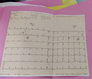
Ecg -ventricular hypertrophy (cardiomyopathy)
The diagnosis may be BECKERS MUSCULAR DYSTROPHY.
ANATOMICAL SITE-MUSCLE
PHYSIOLOGICAL DISABILITY-difficult to squat and change the position from squatting to standing, to climb upstaires, wearing of slippers.
TREATMENT: no cure for this beckers disease yet. Symptomatic treatment, steroids are given to slow down the progression, prednisone may helpful in prduction of utrophin which closely ressembles dystrophin. Orthopaedic surgeons can treat contractures, associated with cardiomyopathy can be treated by ACEinhibitors, ARBS,beta blockers.
Non pharmacollogicaly -physiotherapy, rehabilitation


Comments
Post a Comment